Exercise 21 review sheet gross anatomy of the heart – Embark on a comprehensive exploration of the exercise 21 review sheet on the gross anatomy of the heart, a meticulous guide that unveils the intricate structure and function of this vital organ. Through an engaging narrative, we delve into the external and internal anatomy of the heart, unraveling its chambers, valves, and intricate network of blood vessels.
Prepare to gain a profound understanding of the cardiac conduction system, the heart’s electrical impulses, and the vital role it plays in regulating heart rate and rhythm.
Furthermore, this review sheet illuminates the clinical significance of cardiac anatomy, examining common cardiac diseases and their impact on the heart’s structure. We explore the use of imaging techniques to assess cardiac anatomy and delve into the surgical procedures employed to treat cardiac diseases.
Join us on this captivating journey into the realm of cardiac anatomy, where knowledge and understanding intertwine to empower healthcare professionals and students alike.
Gross Anatomy of the Heart: Exercise 21 Review Sheet Gross Anatomy Of The Heart

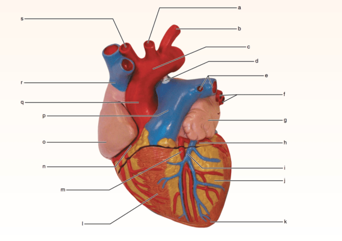
The heart is a muscular organ located in the thoracic cavity, medially in the anterior-inferior portion, slightly shifted to the left. It is surrounded by a double-layered sac called the pericardium, which provides protection, support, and lubrication.
External Anatomy of the Heart
The heart has a conical shape with an apex pointing downwards and slightly to the left, and a base facing upwards and slightly to the right. It is divided into four chambers: two atria and two ventricles. The right atrium and ventricle receive and pump deoxygenated blood, while the left atrium and ventricle receive and pump oxygenated blood.
- Pericardial Layers:The pericardium consists of two layers: the fibrous pericardium (outer layer) and the serous pericardium (inner layer). The serous pericardium is further divided into the parietal pericardium (lining the fibrous pericardium) and the visceral pericardium (covering the heart).
- Major Blood Vessels:The major blood vessels entering the heart are the superior vena cava (SVC) and inferior vena cava (IVC), which bring deoxygenated blood from the body to the right atrium. The pulmonary arteries carry deoxygenated blood from the right ventricle to the lungs for oxygenation.
The pulmonary veins carry oxygenated blood from the lungs to the left atrium. The aorta carries oxygenated blood from the left ventricle to the body.
Internal Anatomy of the Heart
The four chambers of the heart are separated by muscular walls called septa. The atria are separated by the interatrial septum, and the ventricles are separated by the interventricular septum.
- Heart Valves:The heart valves are located between the atria and ventricles, and between the ventricles and the major blood vessels. They prevent the backflow of blood and ensure unidirectional blood flow.
- Coronary Circulation:The coronary arteries supply oxygenated blood to the heart muscle. The coronary veins drain deoxygenated blood from the heart muscle.
Cardiac Conduction System
The cardiac conduction system is responsible for generating and conducting electrical impulses that coordinate the heart’s contractions.
- Components:The cardiac conduction system consists of the sinoatrial node (SA node), the atrioventricular node (AV node), the bundle of His, and the Purkinje fibers.
- Electrical Impulses:Electrical impulses originate in the SA node, which is located in the right atrium. The impulses travel through the atria, causing them to contract. The impulses then reach the AV node, which delays the impulses slightly before sending them to the bundle of His.
The bundle of His divides into the left and right bundle branches, which carry the impulses to the Purkinje fibers. The Purkinje fibers distribute the impulses to the ventricles, causing them to contract.
Blood Supply and Innervation of the Heart, Exercise 21 review sheet gross anatomy of the heart
The coronary arteries supply oxygenated blood to the heart muscle.
- Coronary Arteries:The left coronary artery arises from the aorta and supplies blood to the left atrium, left ventricle, and interventricular septum. The right coronary artery arises from the aorta and supplies blood to the right atrium, right ventricle, and posterior wall of the left ventricle.
- Venous Drainage:The coronary veins drain deoxygenated blood from the heart muscle into the coronary sinus, which empties into the right atrium.
- Innervation:The heart is innervated by the autonomic nervous system, which regulates heart rate and rhythm.
FAQ Section
What are the four chambers of the heart?
The four chambers of the heart are the right atrium, right ventricle, left atrium, and left ventricle.
What is the function of the heart valves?
The heart valves prevent the backflow of blood within the heart, ensuring proper blood flow through the chambers.
What is the significance of the cardiac conduction system?
The cardiac conduction system generates and transmits electrical impulses that coordinate the contraction of the heart chambers, ensuring a regular and efficient heartbeat.